Skin Tabs, Enlarged Papillae and Cryptitis
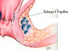
Skin Tabs/TagsEnlarged Papillae
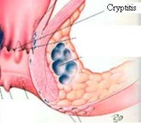
Cryptitis
Anal skin tabs/tags are the shapeless lumps and flaps of skin or flesh found at the anal verge. Anal skin tags are an extremely common condition and are frequently associated with other anorectal problems.
Anal skin tags are usually the result of a prior anorectal insult or injury. An acute swelling of an external hemorrhoid, if left untreated, frequently leaves behind a skin tab - also referred to as a hemorrhoidal tab.

The skin tab's blood supply from the hemorrhoidal artery above may then give rise to the development of an even larger hemorrhoid. Swollen skin edges as a result of prior rectal surgery may also develop into skin tabs.
A sentinel tag is that skin tab which is situated at the inferior border of an infection or injury, as if it is watching or guarding over. A sentry or sentinel is one that keeps guard - thus the name. Anal fissures and fistula are often associated with secondary changes, which may include a sentinel tag. The proximal end of a fissure or fistula may contain granulation tissue that extrudes, beginning the formation of a sentinel tag.
The skin tag is often first noticed by the patient, as a painless soft protrusion beginning near the opening of the anus. If a skin tag is perceived by the patient as causing pain, frequently the physician will find an associated rectal condition which is the actual cause of the pain.
Cleanliness can be a problem. Fecal debris may become trapped under the tag upon wiping in one direction. If there is more than one tab, the problem is multiplied. Itching (pruritus) often develops to make a bad situation worse. Skin tabs may indicate the presence of a more serious rectal ailment that needs careful attention.
Anal tags are generally asymptotic and often are the remnants of previous inflammatory lesion in the anal area. When tags are symptomatic, as a result of itching, pain, anxiety or hygienic problems, they can be removed, and/or biopsied to confirm their etiology. Anoscopy may enable the physician to identify the cause or find other lesions. If tags are small, local anesthetic is injected, then the area is excised. Laser has been used successfully to obliterate skin tabs and resurface the anal area to achieve a good cosmetic result. If extensive, skin tab surgery may need to be undertaken in the operating room. Any surgery in the anal area, no matter how small, may cause some postoperative pain.
Anal papillae are prominent projections of epithelium at the upper end of the anal canal at the mucocutaneous junction. Usually they are small, but visible enough to give the pectinate line a serrated appearance on anoscopy.
Papillae are normal structures causing no symptoms unless they grow large or become inflamed.

They are covered with skin-usually pale pink or whitish-and have a broad base and a fibrous tip. Enlarged papillae may elongate and prolapse at the anal opening during defecation and may need to be replaced digitally.
Papillae may become painful, and reddened. Inflammation of papillae or crypts is frequently associated with fissures, fistulas, Crohn's disease, pruritus ani, and/or internal hemorrhoids. Inflammation of papillae may result from trauma or chemical irritation, such as the passage of hard stools or of irritating liquid stool.
Signs and symptoms of enlarged papillae may be anal discomfort, itching, burning, and sometimes pain-all intensified at bowel movement. An urgent and distressing sensation may occur (tenesmus), as if a discharge from the intestines must take place, although none can be effected.
Papillae must be differentiated from polyps, which they resemble. A biopsy may be helpful in this regard. Polyps are covered by mucosa, may bleed, and are not painful. Papillae are covered by skin, do not bleed, can be painful, and may protrude from the anus if enlarged. Polyps are often pre-cancerous, whereas papillae are not.
Although enlarged papillae can be palpated by the examiner, they are best evaluated on anoscopy. Inflammation or a pustular discharged from an adjacent crypt should be further evaluated to rule out an abscess or fistula.
Treatment should be directed to the underlying condition since most often papillae are secondary to other inflammatory anorectal lesions. Symptomatic papillae are removed by excision. If enlarged anal papillae are associated with internal hemorrhoids, they should be removed as part of the hemorrhoidectomy.
CryptitisAnal crypts are tiny recesses of epithelium at the upper end of the anal canal at the mucocutaneous junction. They are tiny mucus glands of lubrication arranged in a circle around the upper end of the anal canal. Located between normal structures called anal papillae, crypts are usually small, but Cryptitis visible enough to help give the pectinate line a serrated appearance on anoscopy.
Crypts are normal structures causing no symptoms unless they become inflamed. They are small areas of skin situated between the anal papillae. They are approximately 3 mm in depth and are lined with a single layer of epithelium, which is a continuation of the skin of the anus. Just before a bowel movement, the sphincter muscles contract and squeeze out a little drop of lubricating mucus from each of these crypts, aiding in the normal slippery passage of stool.

Cryptitis is held responsible for a variety of conditions and symptoms. The pain of cryptitis is usually of the sharp lancinating or burning variety. A dull ache, or intense pain from spasm of the contraction of the sphincter muscle may develop from the inflammatory process. The nature of a crypt infection is of an ebb and flow, and may be of such a low grade that the pain is transitory.
The cause of cryptitis may be due to an inflammatory process in the adjacent areas, or a disturbance in the acid pH balance of the rectum. Trauma from constipated stools, infections introduced from external sources, parasites, foreign debris, etc., may also initiate cryptitis.Surgical removal of a crypt is not the complete answer to treating cryptitis. The cause must be eliminated.


